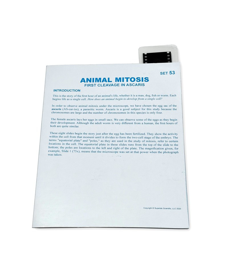
This Microslide, Animal Mitosis, includes a slide featuring 8 related 35mm images photographed through a microscope called photomicrographs. Arrows and call outs help the student locate important features. This Microslide is accompanied by a detailed lesson plan designed to stimulate, inform and question students about the topic under study, and has a pocket in which the Microslide is stored. The following magnified specimens are depicted on the slide: The Zygote (750x), Pro-metaphase (750x), Metaphase (750x), Metaphase-polar view (750x), Early Anaphase (750X), Anaphase (1000X), Telophase (750X), Late Telophase (1000X).
- Simply insert the slide and focus with the Microslide Viewer (sold separately) which is designed for use with Microslides
- Perfect for classrooms or home studies!
- Includes a 35mm slides with 8 magnified specimens


